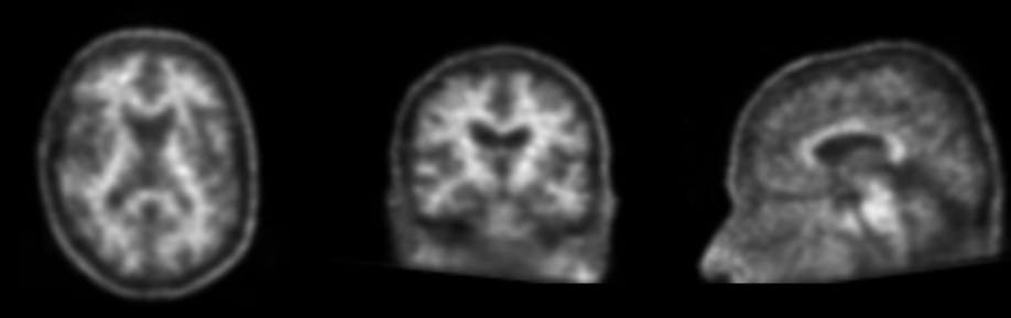
The first step to perform visual assessment accurately, is the orientation or alignment of the PET image.
There are three main hallmarks when properly orientating an amyloid-PET scan:
- first, you need to make sure the midline of the brain is at a vertical axis using the axial orientation;
- second, using the sagittal orientation, you must orient the scan so that the anterior and posterior corpus callosum are on one horizontal line; and
- finally, you need to align the temporal lobes on the horizontal plane using the coronal viewing of the scan, making sure the pons is straight in the middle.
This orientation method is identical for each radiotracer used.
After proper orientation of your scan, the next step is to put it in the correct color scheme and scale to a reference value.
Correct scaling allows you to know what the signal is of the white matter and what the signal should be for the gray matter if there is amyloid in the brain.
Flutemetamol
When you use the flutemetamol radiotracer, this entails putting the color scale to rainbow and scaling the image to 90% intensity of the pons.
This means that the pons should be red or ‘hot’ and this visualization should be apparent with the slide bar at 90% of intensity.
Florbetaben and florbetapir
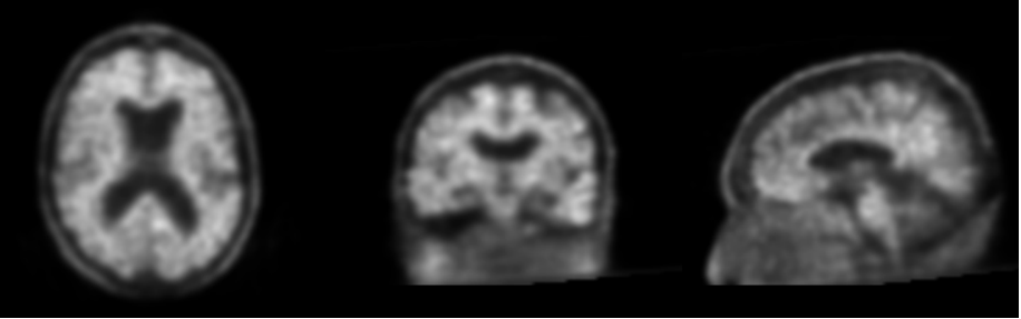
When you use the florbetaben and florbetapir radiotracers, this entails using a grayscale and scaling the image to 90% intensity of the deep white matter.
This means that the deep white matter should be ‘hot’ white.
Flutemetamol
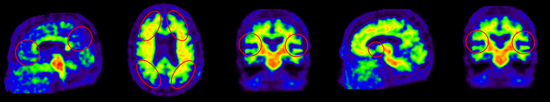
For flutemetamol you assess 5 regions of interest in each hemisphere, so 10 overall, namely: the frontal cortex, lateral temporal cortex, lateral parietal cortex, precuneus, and striatum.
The striatum can only be assessed using flutemetamol.
If one of these regions unilaterally shows increased signal uptake, this is considered to be a positive amyloid-PET scan.
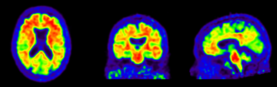
Positive scan

Negative scan
Flutemetamol visual read guidelines
Regions of interest (ROI)
Florbetaben and florbetapir
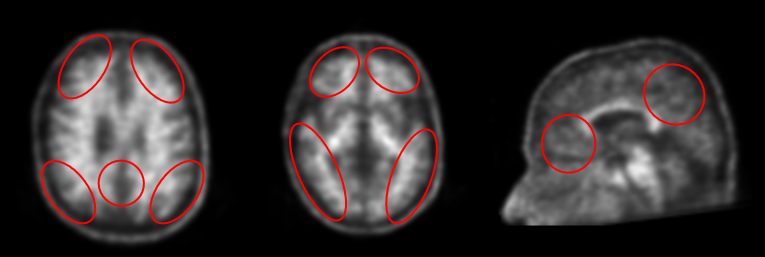
For florbetaben (and also florbetapir, which is assessed in a very similar way only in the inverted gray-scale), you assess 4 regions of interest in each hemisphere, so 8 overall, namely: the frontal cortex, lateral temporal cortex, lateral parietal cortex, and precuneus, but not the striatum.
If one of these regions unilaterally shows increased signal uptake, this is considered to be a positive amyloid-PET scan.
Positive scan
Negative scan
Florbetapir visual read guidelines
Florbetaben visual read guidelines
Regions of interest (ROI)